Your optometrist has many ways to identify issues with your eyes, including vision problems and eye diseases. They use technology during your eye exam to help identify and treat a range of eye conditions. An essential part of your eye exam is an eye health evaluation that involves retinal imaging.
Retinal imaging can help detect many eye conditions, including macular degeneration, diabetic eye disease, glaucoma, and retinal detachment. Performing tests for these conditions can help protect your eye health and overall wellness.
What Is Retinal Imaging?
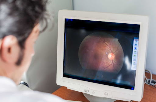
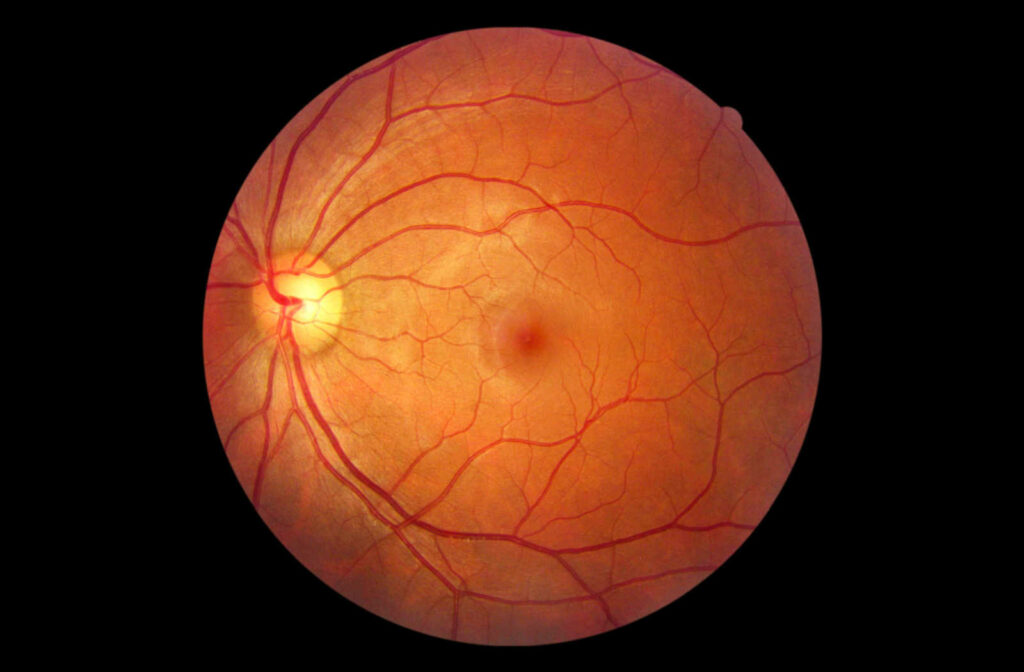
Retinal imaging helps your eye doctor see the inside of your eye in detail. Essentially, your optometrist takes a photo of the internal structures of your eye, including the retina, optic nerve, macula, and surrounding blood vessels. The image your optometrist takes helps them diagnose potential problems like eye diseases.
Depending on your needs, your eye doctor may use different types of retinal imaging.
Why Do You Need Retinal Imaging?
Retinal imaging is a part of many standard comprehensive eye exams. It’s essential for getting an in-depth look at the structures of your eye. Even if you can see well, your eyes may still be developing new conditions—many eye diseases develop with limited symptoms.
These conditions may not show symptoms until they affect your vision. Booking regular eye exams can help protect your eye health.
Retinal imaging lets your eye doctor see the inside of your eye clearly and effectively, helping them identify eye disease as early as possible. The sooner they identify an issue, the faster they can recommend a treatment plan to protect your vision.
What Can Retinal Imaging Detect?
Retinal imaging can detect many eye diseases that go unnoticed by the naked eye. This technology captures detailed images that help your optometrist identify problems they may not notice otherwise.
Some of the conditions and diseases retinal imaging can help detect include age-related macular degeneration, diabetic eye disease, glaucoma, and retinal detachment.
Age-Related Macular Degeneration
Age-related macular degeneration (AMD) occurs when the macula, the part of your eye responsible for central vision, begins to thin. This condition can develop naturally with age, affecting your ability to see what’s directly ahead of you.
There are 2 forms of AMD: wet and dry. Treatment for this disease depends on the kind you have. Individuals with AMD typically develop the dry form first, which can progress to the wet form.
Diabetic Eye Disease
Diabetic eye disease is a general term to describe eye problems that develop because of diabetes. In general, diabetes increases your risk of several eye conditions, including glaucoma and cataracts.
One disease specific to diabetes is diabetic retinopathy, which occurs when blood vessels in your retina, the thin layer of tissue in your eye responsible for sending signals to your brain, begin to swell and leak into the eye.
High blood sugar can cause issues with the blood vessels in the retina, causing fluid and blood to leak. As this disease progresses, it can lead to vision loss, scarring, and other complications.
Glaucoma
Glaucoma is a group of eye diseases that damage your optic nerve, an essential part of your eye necessary for sight. Many forms of glaucoma increase intraocular pressure (IOP), damaging the optic nerve. With time, this disease can lead to severe vision loss.
Glaucoma is a leading cause of blindness in adults over 60, making regular eye exams essential for protecting your vision. Depending on its form, glaucoma may not present many symptoms until vision loss begins. Your optometrist uses retinal imaging during your eye exam to identify early signs of glaucoma.
Common types of glaucoma include:
Retinal Detachment & Tears
Retinal detachment is an emergency. It occurs when your retina pulls from its natural position in the eye. Detachment removes the retina from oxygen, increasing your risk of vision loss the longer the eye goes untreated.
Your eye doctor can diagnose retinal detachment by completing a comprehensive eye exam. They look at the back of your eye to identify holes or tears in the retina.
There are several signs you may have a detached retina:
- The sudden appearance of floaters in your vision
- Blurry vision
- Reduced peripheral vision
- A shadow over your visual field
- Flashes of light
Eye Cancer
Cancer can either start in the eye or spread to the eye from another part of the body. The most common type of eye cancer is intraocular melanoma, cancer that forms inside the eye.
While cancer can develop in the conjunctiva, a clear tissue covering the inside of your eyelid, it more commonly develops in the uvea. The uvea is the middle of your eye, and cancer can form in the iris, the muscles in the eye, or near the retina.
Your optometrist can use retinal imaging to help identify early signs of cancer.
Damage from High Blood Pressure
High blood pressure can negatively affect your health, including your eyes. This problem typically occurs due to diabetes, high cholesterol, or smoking. Thankfully, retinal imaging can help catch this damage.
High blood pressure can damage blood vessels in your retina and increase your risk of other complications. You’re at more risk of damage the longer you have high blood pressure.
Protect Your Eye Health & Vision
Your vision is precious, and retinal imaging is essential for protecting your sight. It’s one of the many tools your optometrist has to care for your eye health and vision. Book your next eye exam, and your eye doctor can complete a detailed examination.
Contact Park Slope Eye when it’s time for your next eye exam.